

2D-ICE
Delivers High-Resolution Imaging for Precise and Safe Cardiac Interventions
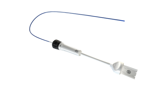
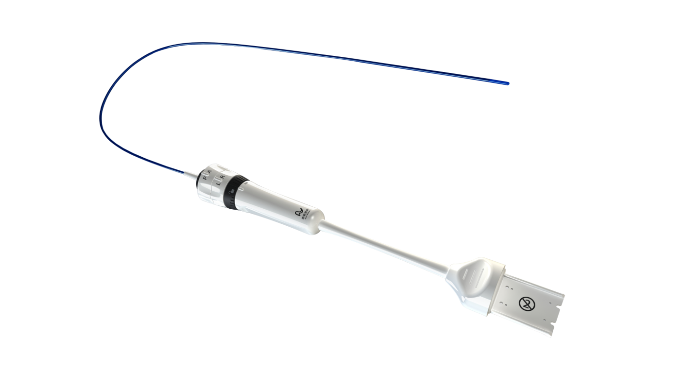
Intracardiac echocardiography (ICE) is an imaging technique that uses a small high-frequency ultrasound probe placed inside the heart to capture clear, high-resolution images. It helps guide procedures like arrhythmia ablation, left atrial appendage closure, and valve replacement by providing detailed views of heart structures and blood flow.
2D-ICE
Delivers High-Resolution Imaging for Precise and Safe Cardiac Interventions
Intracardiac echocardiography (ICE) is an imaging technique that uses a small high-frequency ultrasound probe placed inside the heart to capture clear, high-resolution images. It helps guide procedures like arrhythmia ablation, left atrial appendage closure, and valve replacement by providing detailed views of heart structures and blood flow.
-

High-Resolution Imaging
Provides detailed images of heart chambers, valves, and blood flow for accurate diagnosis and real-time evaluation of treatment
-

Precise Interventional
Provides real-time imaging of heart chambers, valves, and blood flow for accurate guidance during interventions.
-
Enhanced Patient Comfort
Improves patient comfort and efficiency by avoiding radiation and general anesthesia.
-
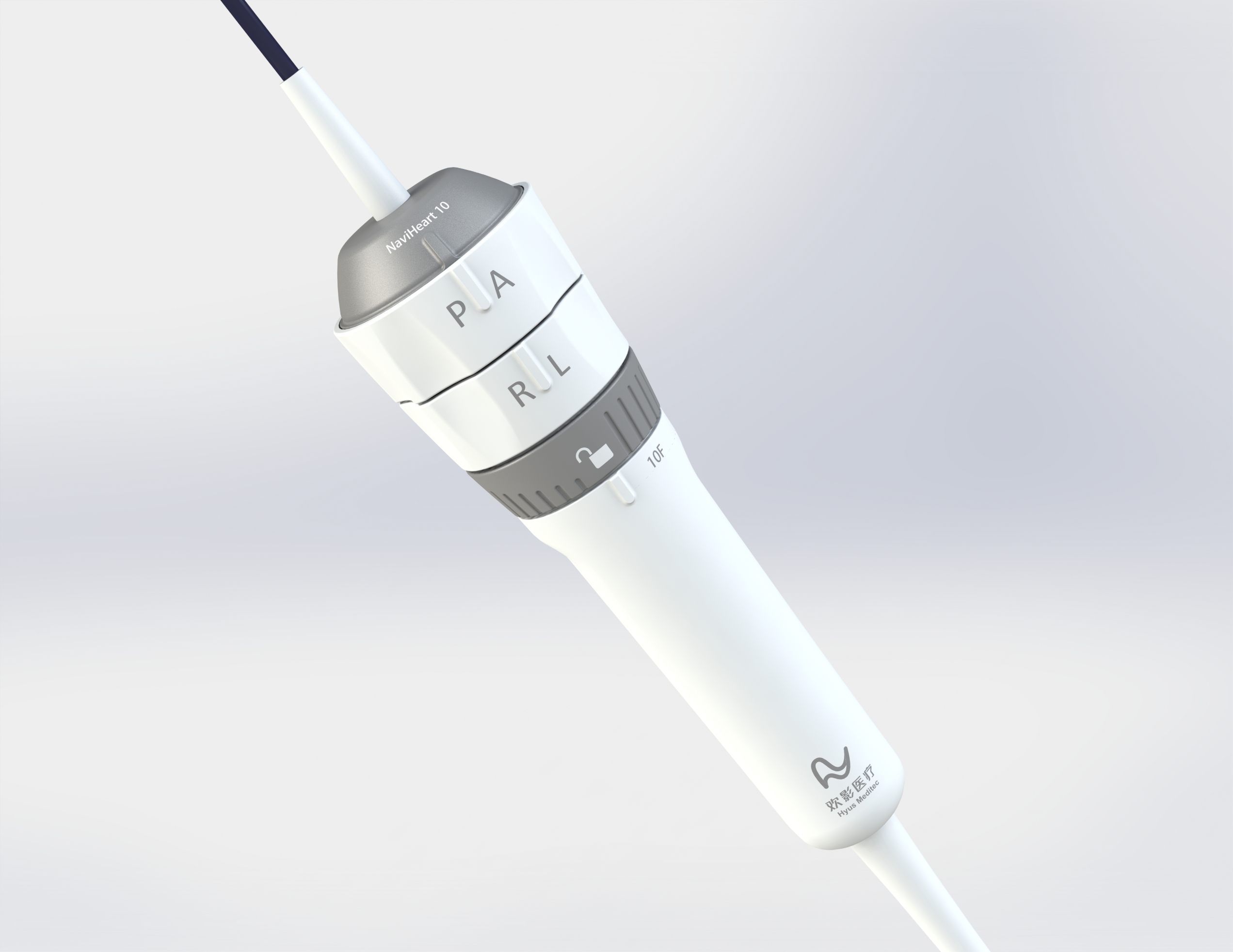
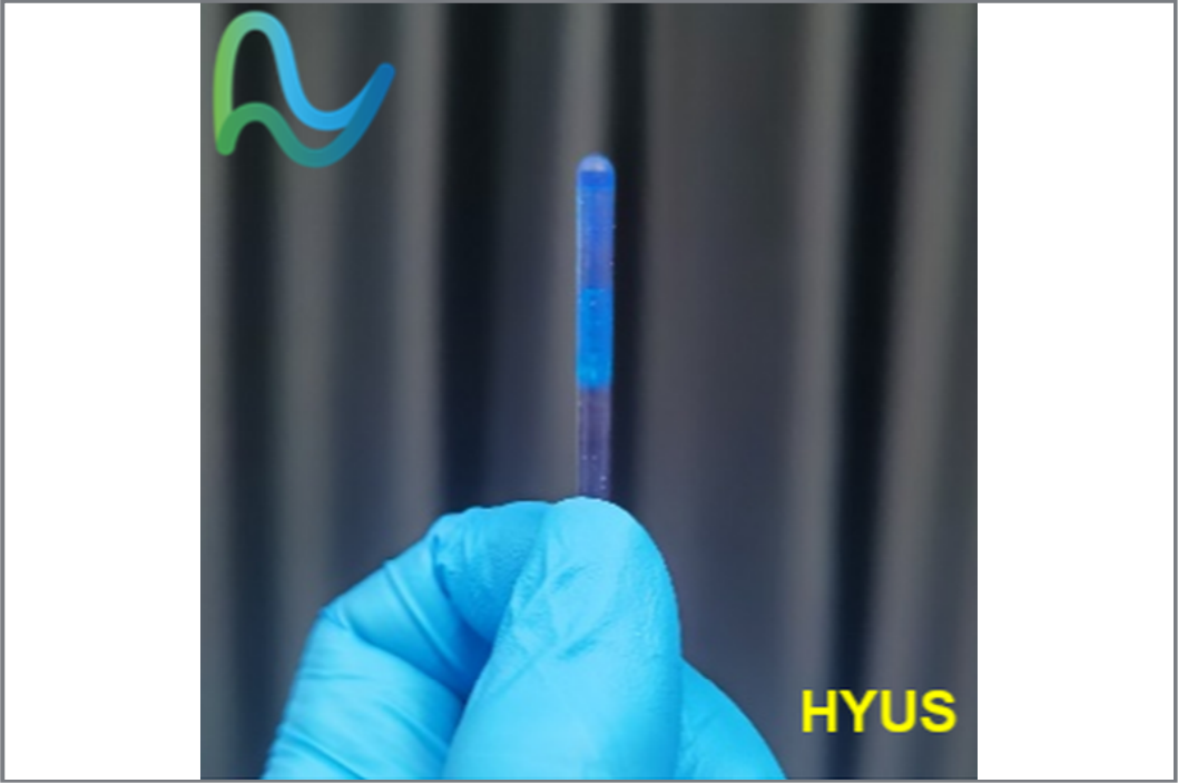
Variety of Catheter Models
Multiple catheter sizes (8F, 10F) to suit different anatomies and surgical needs.
- A. Enriquez, et al, “Use of Intracardiac Echocardiography in Interventional Cardiology Working With the Anatomy Rather Than Fighting It”, Circulation, vol. 137, no. 21, pp. 2278-94, May 2018.
- J. Zhong, et al, “Intracardiac echocardiography Chinese expert consensus”, Front. Cardiovasc. Med., vol 9, no. 1012731, 2022.