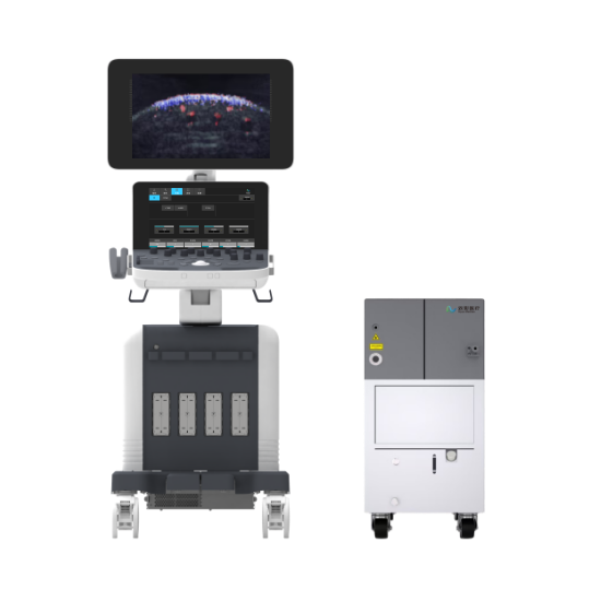


Sonorover UR820
High Resolution Imaging System for Small Animal Studies
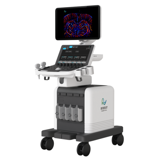
The Sonorover UR820 is an ultra-high frequency ultrasound system for small animals, offering flexible probes and high-resolution imaging. It supports advanced functions like contrast, strain, and super-resolution imaging, meeting research needs in cardiovascular, abdominal, tumor, and embryo studies.
Sonorover UR820
High Resolution Imaging System for Small Animal Studies
The Sonorover UR820 is an ultra-high frequency ultrasound system for small animals, offering flexible probes and high-resolution imaging. It supports advanced functions like contrast, strain, and super-resolution imaging, meeting research needs in cardiovascular, abdominal, tumor, and embryo studies.
-

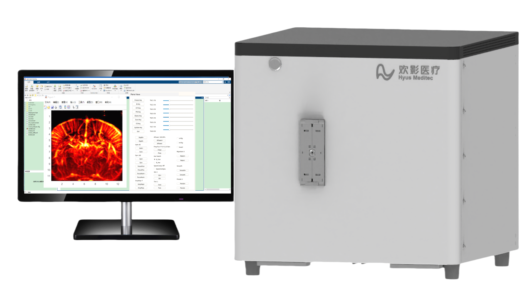
Advanced Imaging Platform
With GPU-accelerated processing and powerful computing for advanced imaging algorithms.
-
256-Channel
The 256-channel ultrasound system provides clearer images with higher resolution and better signal quality to show tissue and pathology details.
-
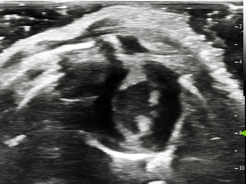
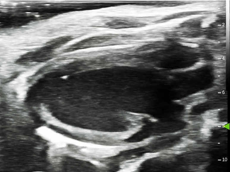
Ultra-High Resolution
The system provides clear images of tiny structures and fine anatomy for accurate diagnosis.
-
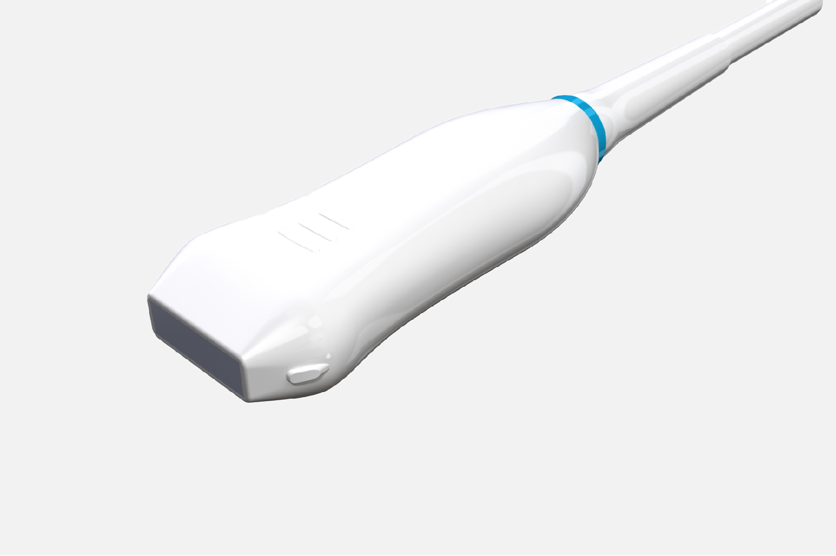
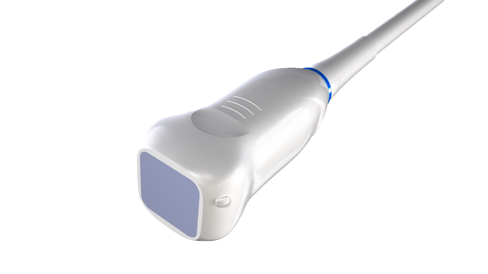
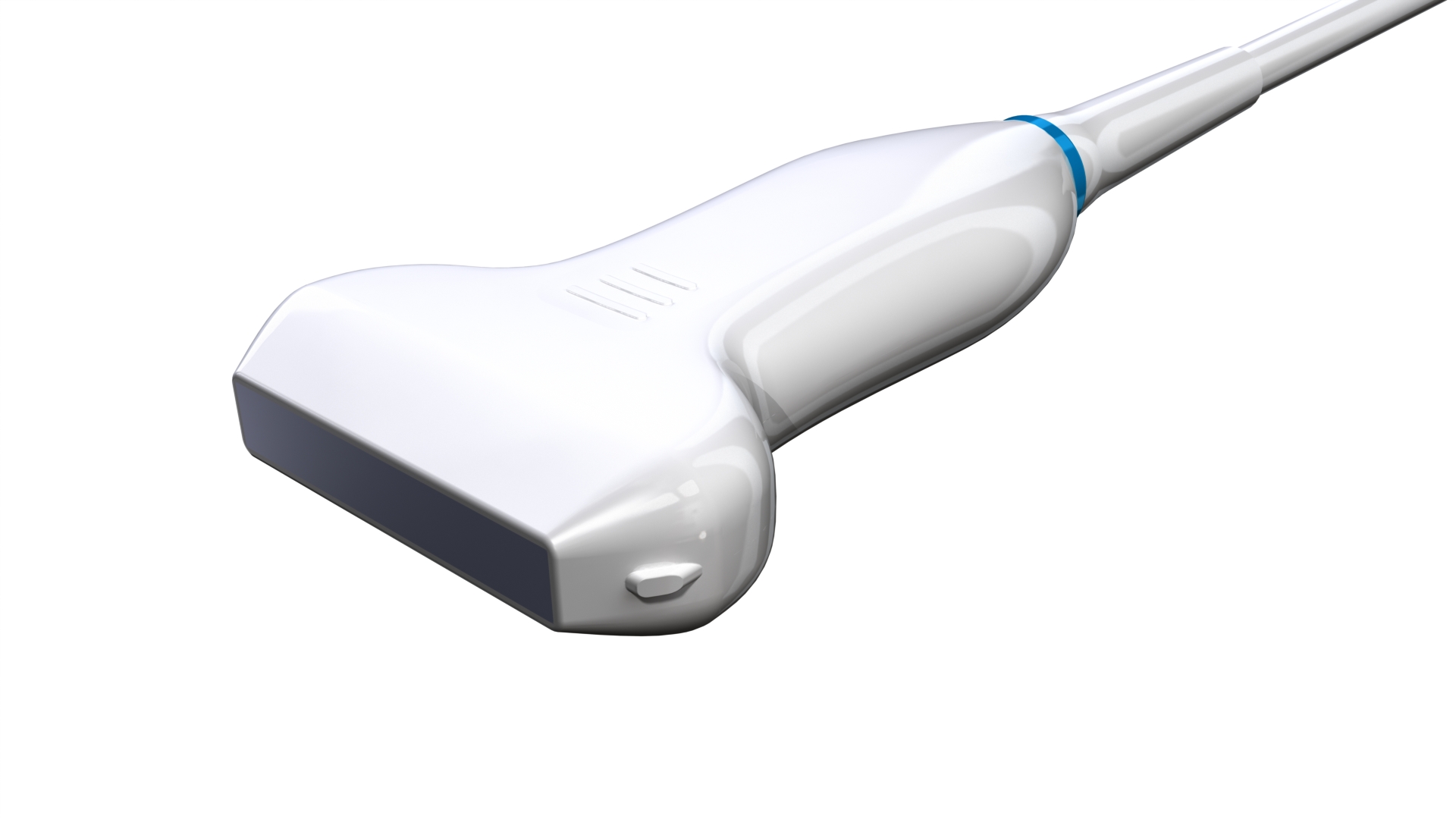
High-Frequency Probes
Offers flexible probe options to cover imaging needs for large and small animals, including heart, abdomen, tumors, and embryo studies.
-
Super-Resolution Ultrasound Imaging
Utilizing microbubble localization and tracking to achieve sub-wavelength imaging, overcoming the diffraction limit of conventional ultrasound. Breaks spatial resolution barriers to visualize microvascular network details, providing new perspectives and data support for studying tissue microstructures and evaluating blood flow characteristics.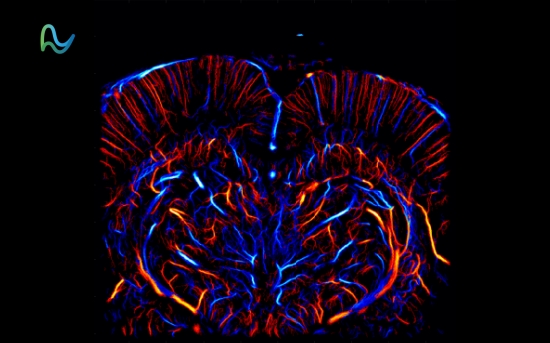
-
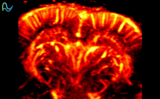
Micro Flow Imaging
Captures low-speed, micro-scale blood flow signals to visualize fine vascular networks and blood flow dynamics. Applications include rat/mouse whole-brain blood flow imaging, early lesion detection, tumor characterization, and tissue perfusion monitoring. -
Myocardial Strain Imaging
Accurately assesses cardiac function through quantitative analysis of myocardial strain parameters, enabling detection of subtle myocardial abnormalities and providing reliable data for cardiovascular research and diagnosis.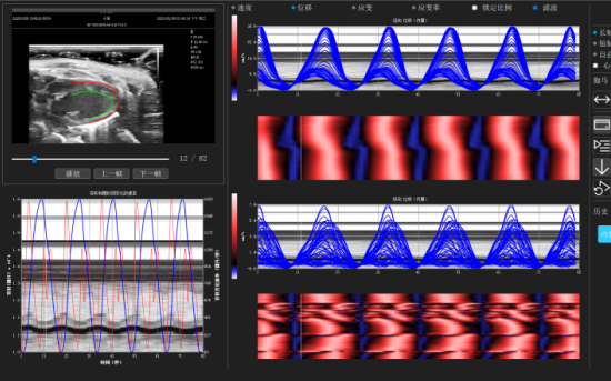
-
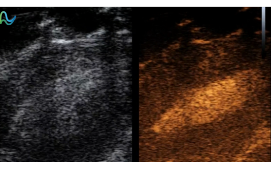
High-Frame-Rate Contrast Imaging
Achieves superior temporal resolution by increasing imaging frame rate, enabling clearer visualization of microvascular architecture and perfusion details. -
Photoacoustic/Ultrasound Dual-Modality Imaging
Combines functional imaging via laser-induced ultrasound signals with structural imaging via high-frequency ultrasound echoes. The photoacoustic module supports real-time imaging up to 50 fps, precisely capturing hemodynamic processes and enabling accurate correlation of functional and structural information.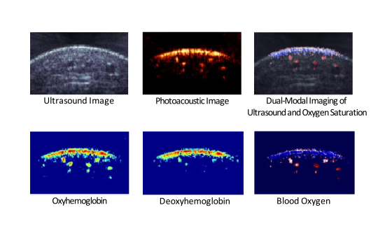
-
Mouse Heart Left Ventricle Long-Axis View
-
Mouse Heart Left Ventricle Short-Axis View
-
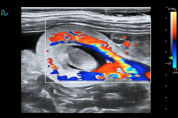
Rat Heart Left Ventricle Short-Axis View
-
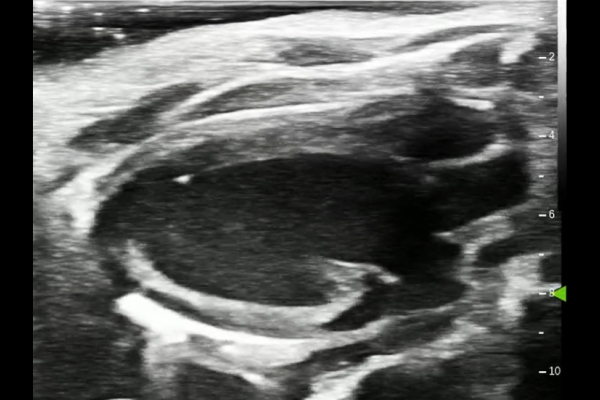
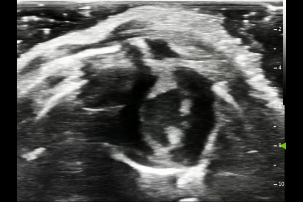
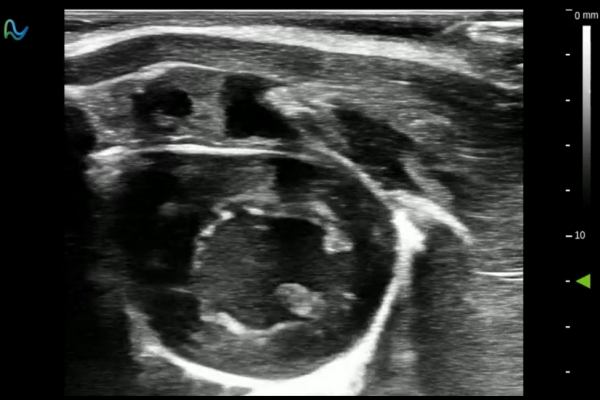
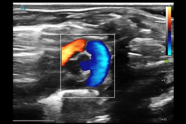
Rat Kidney Blood Flow
-
Rat Heart Aortic Blood Flow
-
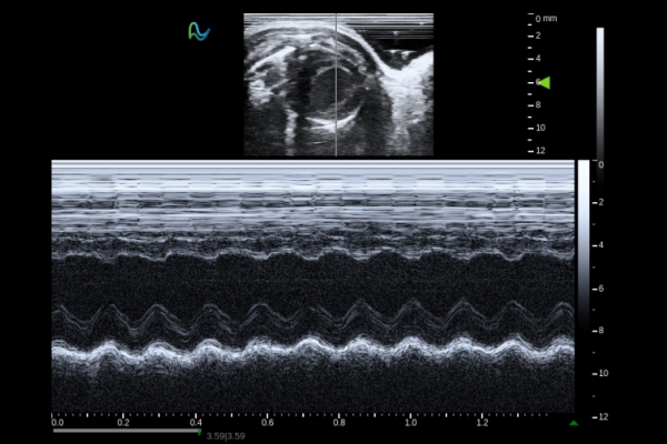
Mouse Heart Left Ventricle Short-Axis M-mode
-
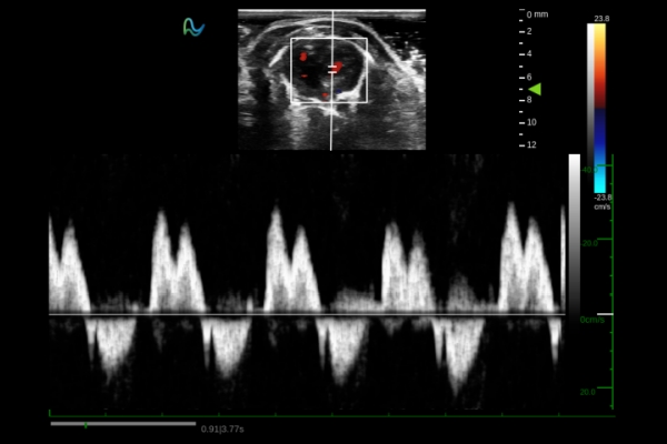
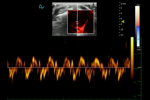
Rat Heart Apical Four-Chamber PW Mode
-
Mouse Heart Apical Four-Chamber TDI Mode
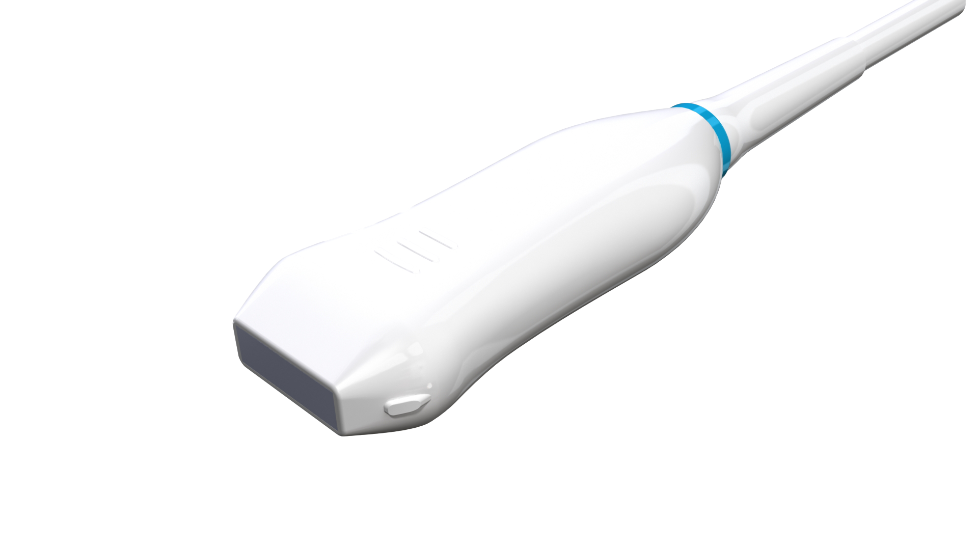
L42-18A
Application: Zebrafish heart, Mouse cardiovascular, abdomen, and embryos.
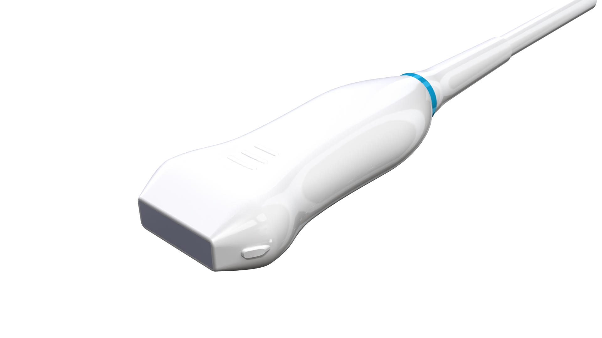
L32-14A
Applications: Rat (<250g) heart and abdomen.
L21-9A
Applications: Rat (>250g) heart and abdomen.
L14-6A
Applications: Rabbit heart and abdomen.
L12-4A
Applications: Rabbit heart and abdomen.